facebook
ClearView Logo
Home   •    Patient Resources   •    Blog

What Is Retinal Detachment? Types, Signs, Symptoms, and Treatment

retinal detachment
retinal detachment

Retinal detachment is a critical condition of the eye that happens when the retina, the thin layer of tissue at the back of the eye gets detached from its normal place. Such a separation hinders the retina from performing its functions properly. In fact, this can cause irreversible blindness at the same time if treatment is delayed. Learning about the types, signs, symptoms, risk factors, and therapies of retinal detachment is very important for taking care of one's eyes and making sure that you get a doctor consultation without delay.


What Is Retinal Detachment?

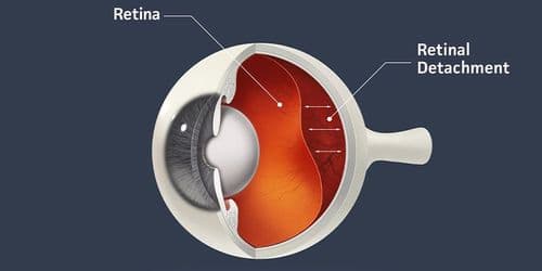
To understand retinal detachment it is important first to understand the role of the retina in the eye. The retina functions by converting light into visual signals that the brain then recognizes as images. If the retina becomes detached such signals will be broken causing vision loss or even total blindness in that eye.

Usually the causes of retinal detachment are trauma, eye surgery history, certain eye diseases, more common in middle-aged or elderly patients. Also, it can occur after a perforating injury or foreign body. It is very important to make a quick diagnosis and start the treatment immediately to save the patient's vision.


Types of Retinal Detachment

To establish an effective treatment plan, it's important to identify the various classifications of retinal detachment. These classifications include the following:

1. Rhegmatogenous retinal detachments.

  • Rhegmatogenous retinal detachments are by far the most encountered type. The cause of rhegmatogenous detachment is due to a defect in the retina which causes fluid from the vitreous (gel-like fluid in the eye) to leak under the retina leading to additional detachment of the retina.
  • Most rhegmatogenous detachments occur in patients over the age of 50 due to shrinkage of the vitreous gel pulling away from the retina, resulting in defect formation.

2. Tractional Retinal Detachment:

  • When the surface of the retina is scarred and contracts by pulling away the retina from its normal position, it is classified as this type of detachment.
  • This type of detachment usually occurs in people with diabetes, particularly those who have developing abnormal blood vessels coupled with diabetic retinopathy.

3. Exudative Retinal Detachment:

  • This type of retinal detachment does not occur as a result of a tear or break in the retina, but rather due to an accumulation of fluid that builds up under the retina, which can occur due to several reasons, including inflammatory diseases, tumors, or vascular disease.
  • Exudative (serous) retinal detachment can be caused by several conditions such as age-related macular degeneration (AMD) or uveitis.

By understanding the different types of retinal detachment, both patients and their health care providers can identify the nature of the retinal detachment, the potential risks associated with it, and what may happen as a result of having exudative retinal detachment.


Signs of Retinal Detachment

Signs of retinal detachment can differ based on the type and severity of the specific detachment (each will have its own signs). The earlier a retinal detachment is recognized, the more favorable the treatment outcome will be. Following are the common signs associated with retinal detachment:

  • Flashes of Light - You may see flashes of light (suddenly appearing) at the edges of your field of vision. This is indicative that the retina is being stimulated (due to the pulling of the vitreous gel from vitreous separation).
  • Floaters - floaters like tiny dots or lines may appear in your vision as the vitreous pulls away from the retina.
  • Dark Shadows or Curtains - A shadow or curtain-like appearance in your field of vision may occur if the retina is detaching.
  • Blurriness - If you notice gradual blurriness or a decrease in visual clarity, this could also be a sign that the retina is becoming detached.


Symptoms of Detached Retina

The symptoms of a detached retina can escalate quickly, and individuals may experience a combination of the following:

  • Sudden Vision Loss: A significant and sudden loss of vision in one eye can occur, which may be partial or complete.
  • Distorted Vision: Straight lines may appear wavy or distorted, indicating that the retina is not functioning correctly.
  • Peripheral Vision Loss: A sudden reduction in side vision can also signify retinal issues.

If you experience any of these symptoms, it's essential to seek medical attention immediately.


Risk Factors for Retinal Detachment

Understanding the risk factors for retinal detachment can aid in prevention and early detection. Some of the primary risk factors include:

  • Age: The risk increases as you age, particularly after 40 years old.
  • Previous Eye Surgery: Individuals who have had cataract surgery or other eye surgeries may be at higher risk.
  • Trauma: Eye injuries or trauma can lead to retinal tears or detachments.
  • Family History: A family history of retinal detachment may increase your risk.
  • High Myopia: Individuals with severe nearsightedness (myopia) are at a higher risk due to the elongated shape of their eyeball.
  • Other Eye Conditions: Conditions such as diabetic retinopathy, uveitis, or retinitis pigmentosa can elevate the risk of detachment.

Being aware of these risk factors can empower individuals to take proactive measures to protect their eye health.


Retinal Detachment Treatments

Timely treatment for retinal detachment is critical for preserving vision. The treatment method depends on the type of detachment and the extent of damage. Common retinal detachment treatments include:

1. Laser Surgery:

  • Laser surgery is often used to treat rhegmatogenous retinal detachment. In this procedure, the ophthalmologist uses a laser to create tiny burns around the retinal tear, contact lenses, LASIK Surgery, and cataract surgery for those with age-related vision issues., sealing it and preventing further fluid from entering the space under the retina.
  • Laser surgery can effectively halt the progression of the detachment and is typically performed on an outpatient basis.

2. Cryopexy:

  • This technique involves freezing the area around the tear, which helps adhere the retina back to the underlying tissue. It is often used in conjunction with laser surgery.

3. Pneumatic Retinopexy:

  • This procedure involves injecting a gas bubble into the eye, which helps push the detached retina back into place. Patients may need to maintain a specific head position for the gas bubble to be effective.

4. Scleral Buckling:

  • In this surgical procedure, a silicone band is placed around the eye to push the wall of the eye against the retina, helping to reattach it. This is often used for more extensive detachments.

5. Vitrectomy:

  • This surgery involves removing the vitreous gel that is pulling on the retina. It may be combined with other procedures to treat more complex retinal detachments.

The choice of treatment will depend on various factors, including the type and extent of the detachment, the patient's overall health, and the ophthalmologist's recommendation.


Conclusion

To sum it up, in order to keep your vision and eye health intact, it is necessary to fully understand all aspects of retinal detachment including what it is, how many types of retinal detachment exist, what the signs/symptoms of retinal detachment will be and what treatments there are for retinal detachments. If detected early, you will have a better chance at a good outcome with any serious condition, so if you experience any flashes, new floaters, or sudden vision loss, please seek medical assistance immediately.

By knowing your risk factors for developing a retinal detachment as well as which types of retinal detachments there are, you can take action to help protect your own vision. Regular eye exams and being aware of changes to your own sight are both essential for maintaining overall eye health.

GET STARTED WITH CLEARVIEW

Schedule your free consultation with Dr. Sandy T. Feldman or Dr. Michael. L Mathison to discover how achieving visual freedom can change your life.

Google Logo
Facebook logo large
Facebook Logo
Yelp Logo